
Adult and Neglected Clubfoot: Understanding, Evaluation and Staged Ilizarov Correction
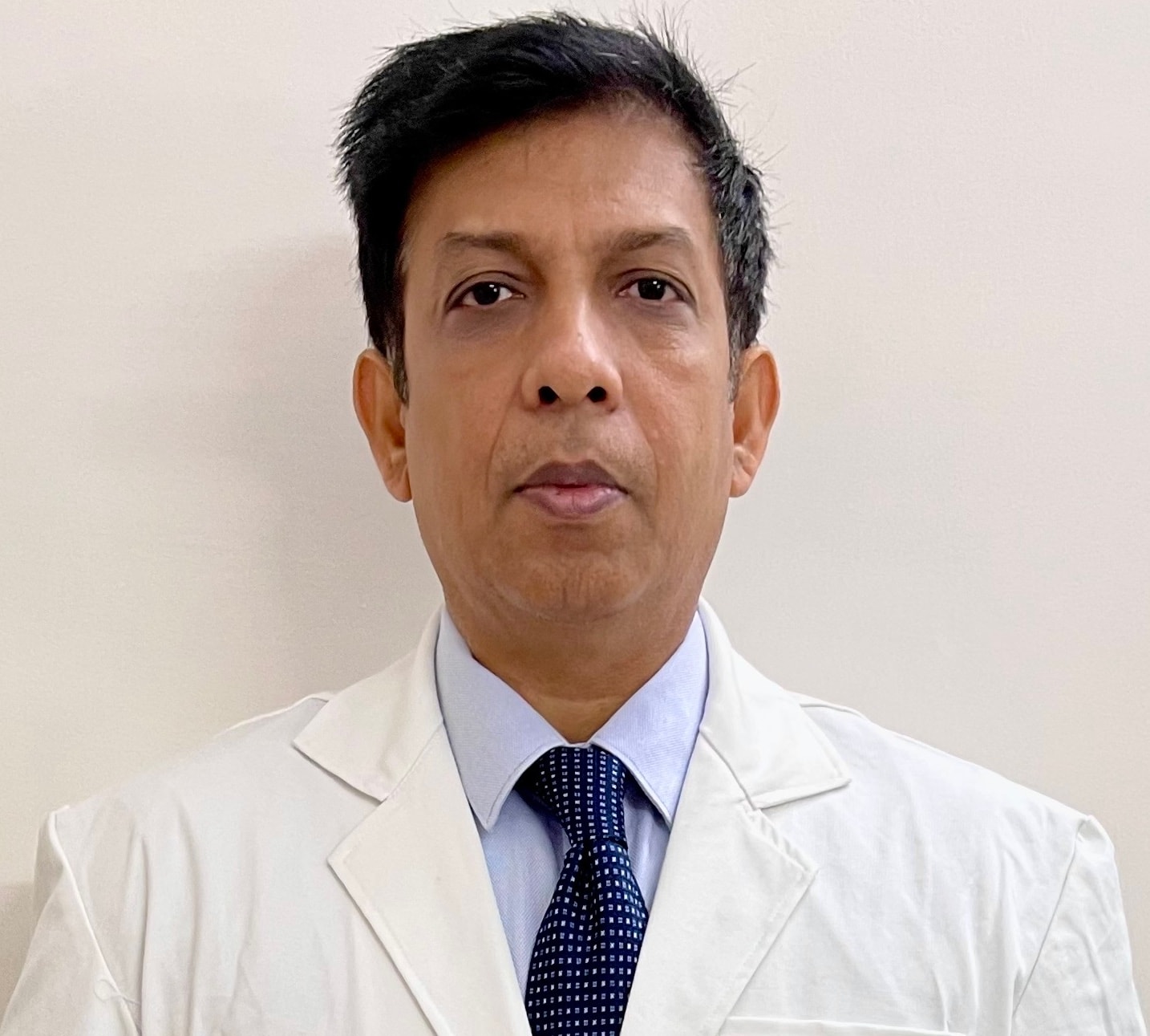
Written and medically reviewed by Dr. Shamsul Hoda, MBBS, D.Ortho., MS Ortho, M.Ch. Orth., Dip. Ilizarov (Kurgan)
Last reviewed: November 2026
Clubfoot is among the most common birth deformities. In many parts of India, Nepal and Bangladesh, children still reach adolescence or adulthood without effective treatment, or the deformity returns after early care. Adults then walk on the outer border or the top of the foot, develop thick calluses and ulcers, cannot wear ordinary shoes, and face social and work difficulties. It is not too late to correct an adult clubfoot. This article explains how we assess and treat it.
Understanding the deformity
Clubfoot has four components, remembered as CAVE:
- Cavus – a high arch from a dropped first metatarsal
- Adductus – the forefoot turned inwards
- Varus – the heel turned inwards
- Equinus – the foot pointed down, with a tight Achilles tendon
In a baby these are mainly soft-tissue contractures and the bones are cartilaginous and adaptable. In an untreated adult, years of walking on a deformed foot reshape the bones: the talus becomes wedge-shaped, joints adapt or stiffen, skin and vessels shorten, and weight-bearing calluses or ulcers form over the outer border and dorsum. That is why adult correction is different from infant correction.
Clinical evaluation
- Standing and walking: which part of the foot bears weight, and the presence of callus or ulcers
- Rigidity of each component – how much corrects passively
- Skin quality, scars from earlier surgery, sensation and pulses
- Calf muscle bulk and strength; tendon balance (especially tibialis anterior and posterior, peronei)
- Associated conditions: arthrogryposis, spina bifida or other neurological causes change the plan and the expected result
- The whole limb: knee, rotation, leg-length difference
- The patient's goals, occupation, footwear and ability to attend follow-up
Radiography and imaging
Weight-bearing AP and lateral X-rays of the foot and ankle are essential. We look at:
- Talocalcaneal angles on AP and lateral views (parallelism of talus and calcaneus is typical in clubfoot)
- Talo–first metatarsal angle (forefoot adductus and cavus)
- Tibiocalcaneal angle (equinus)
- The shape of the talar dome and ankle joint congruity
- Navicular position and midfoot joint changes, arthritis
CT scanning helps with severe bony deformity and surgical planning. The deformity is then planned digitally so that the site and order of correction are clear before surgery.
Treatment options
Ponseti casting. The Ponseti method is the standard of care for infants, and serial casting can also help older children and adolescents with some flexibility. In stiff adult feet it is rarely enough alone but may soften soft tissues before surgery.
Bracing and footwear. Used after correction to hold the result, and for patients who are not candidates for surgery.
Acute surgery. Extensive soft-tissue release, osteotomies or triple arthrodesis can correct moderate deformity in one stage, at the cost of shortening the foot and placing stress on tight skin and vessels.
Gradual correction with the Ilizarov method. A circular frame on the leg and foot allows the deformity to be corrected slowly, a little each day. Because skin, nerves and vessels stretch gradually, large corrections can be achieved through small incisions, often while avoiding or limiting bone removal and preserving foot length.
Ilizarov surgical planning – the staged approach
Stage 1 – Planning. Digital planning on weight-bearing X-rays decides whether correction can be achieved through soft tissues and joints alone, or whether osteotomies are needed (for example, a U- or V-osteotomy through the hindfoot and midfoot, or a midfoot osteotomy). The frame is designed with hinges and motors placed for each component.
Stage 2 – Surgery. A frame is applied with rings on the tibia, a foot plate, and wires through the heel and forefoot. Achilles tendon lengthening, plantar fascia release, and any planned osteotomies are performed. The toes may be protected with wires to prevent clawing.
Stage 3 – Gradual correction (usually several weeks). After a short waiting period, the frame is adjusted daily. The joints are first gently distracted to protect cartilage, then cavus, adductus, varus and equinus are corrected in a planned sequence. Regular X-rays, skin checks and nerve checks guide the speed.
Stage 4 – Holding (consolidation). Once a plantigrade foot is achieved, the frame is kept in place for a period so the new position stabilises; if osteotomies were done, until bone heals.
Stage 5 – Frame removal and casting. The frame is removed under anaesthesia, and a walking cast is applied for some weeks.
Stage 6 – Bracing and rehabilitation. Custom footwear or an ankle-foot orthosis maintains correction while muscles and gait retrain.
Possible complications
- Pin and wire site infection; wire breakage
- Skin tension problems or wound breakdown
- Temporary nerve stretch symptoms
- Claw toes from tight toe flexors (prevented or treated with wires or tenotomy)
- Stiffness – an adult corrected foot is usually stiffer than a normal foot
- Under- or over-correction, or a rocker-bottom shape if the midfoot breaks instead of the hindfoot correcting
- Recurrence, especially in neurological feet or if bracing is not followed
- Pain from pre-existing arthritis
Rehabilitation protocol (typical)
- During the frame: weight-bearing as advised, daily pin care, toe and knee exercises, elevation
- After frame removal: walking cast, then gradual weight-bearing in a boot
- After casting: custom shoes or brace, gait training, calf and foot strengthening, balance work
- Long term: footwear review and annual check-ups, sooner if callus, pain or recurrence appears
Long-term outcome
The goal is a plantigrade, painless foot that fits in a shoe, with healed skin and a better walking pattern. Most adults gain a far more functional foot, but it will not be a normal foot: some stiffness and shoe modification are common, and neurological feet need closer monitoring. We discuss realistic expectations before surgery.
For doctors: Adult neglected clubfoot is a specialist reconstruction. Please refer with weight-bearing AP and lateral foot and ankle radiographs, photographs of the weight-bearing area and callus, and history of previous treatment.
This article is for patient education and does not replace a consultation. Treatment and results differ for every patient.