
Congenital Pseudarthrosis of the Tibia: What Parents Should Know
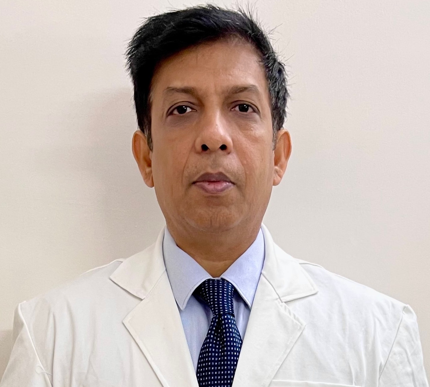
Written and medically reviewed by Dr. Shamsul Hoda, MBBS, D.Ortho., MS Ortho, M.Ch. Orth., Dip. Ilizarov (Kurgan)
Last reviewed: November 2026
Congenital pseudarthrosis of the tibia (CPT) is a rare condition in which the shin bone is weak from birth at one spot. The leg often shows a forward-and-outward bow in infancy. The weak area tends to break, and once broken it often fails to heal – forming a "false joint" (pseudarthrosis). It is one of the most challenging problems in children's orthopaedics, but with modern techniques most children can achieve a healed, functional leg.
Who gets it
About half of affected children have neurofibromatosis type 1 (NF1) – look for light-brown "café-au-lait" skin patches. Others have no associated condition. A child with CPT should be checked for NF1, and a child with NF1 and a bowed shin should be seen early.
Stages
- Before fracture: the tibia is bowed and narrowed or cystic, but intact
- After fracture: the bone has broken and is not healing, often with tapering bone ends and an abnormal thickened sleeve of tissue around the site
The most important rule: protect the bowed leg
A bowed tibia in CPT must not be straightened by a routine osteotomy, because cutting the bone can create a pseudarthrosis that will not heal. Before a fracture, the aim is to prevent it: a protective brace (clamshell orthosis or knee-ankle-foot orthosis) is worn whenever the child is up and about.
Treating an established pseudarthrosis
Modern treatment combines several principles at one operation:
- Removal of the abnormal tissue – the diseased bone ends and the thickened sleeve of tissue around them
- Bone grafting – a generous graft, often from the pelvis
- Stable fixation – commonly a rod inside the tibia, with an Ilizarov frame for compression, alignment and, when needed, bone transport or lengthening
- Strategies to improve union – such as creating a bony bridge between tibia and fibula (cross-union) in selected cases
Vascularised fibula transfer is another option in some centres. When repeated surgery has failed, amputation with a good prosthesis is sometimes the best route to an active life – this is discussed openly with families, never as a first choice.
After the bone heals
- Bracing until skeletal maturity – refracture is the main long-term risk
- Regular X-rays during growth
- Monitoring for leg-length difference and ankle valgus, which can be treated with guided growth or lengthening later
Possible complications
Refracture, failure to unite, leg-length difference, ankle deformity, stiffness, pin-site problems, and the need for further surgery. CPT needs a long-term partnership between family and surgeon.
For doctors: Please avoid corrective osteotomy for anterolateral tibial bowing in infancy. Refer early with full-length tibia radiographs and note any café-au-lait patches.
This article is for patient education and does not replace a consultation. Treatment and results differ for every patient.